EDPA Case of the Month
The EDPA Case of the Month is graciously provided by Dr. Richard Miller. All comments, suggestions and approaches are the opinion of Dr. Richard Miller. The EDPA is not liable for the content, animals, procedures or opinions of the following cases provided.
Biographical Information: Richard Miller graduated with highest honors from the Washington State School of Veterinary Medicine in 1961. For the next fifteen years, he practiced in Lexington, Kentucky, in a primarily reproductive and pediatric practice with a good mix of sport horse issues, as he was also involved in polo and fox hunting. He operated Summerhill Farm in Lexington, as a combination breeding, foaling and training program, before moving to Southern California to join the Equine Medical Center in Cypress, Ca. The next eleven years were occupied by an exclusive racetrack practice at Los Alamitos and Bay Meadow. In 1987, Dr. Miller decided to retire from the track and dedicate his practice to equine dentistry.
For the last 10 years, he has served an area from San Diego to Sacramento, with a 28 ft. mobile clinic that is a full service unit, staffed by him and two technicians. He is an EDPA Certified Equine Dental Provider, advanced certified by the International Association of Equine Dentistry.
The full collection of "Case of the Month" NOW available in book form!
Miller's Digest of Equine Dental Cases can be purchased through the EDPA. Click here to order yours today!
~Case of the Month~
Case of the Month:
Cause and Affect
By: Richard O. Miller DVM, EDPA/C
September 2017
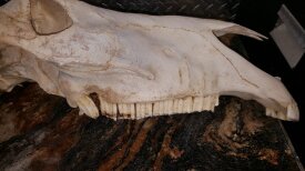
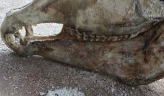
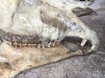
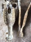
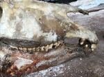
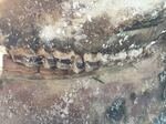
This skull demonstrates several abnormalities, malocclusions and asymmetries, especially in the mandible but I would rather point out another issue that is often discussed at conferences and workshops – the relationship between diagonal bites and molar table angle. Clinically, I seldom see the relationship but with this severe diagonal and the obvious difference in molar table angle, it is suggested that the diagonal affects the preferred side of mastication. In this case, it appears that mastication occurs principally on the 100 side. Basic physics and the chewing cycle would suggest that this is the case but arguments to the contrary are invited.
To see more photos of this case and comments from the Michigan State University Veterinary School- come to the 2017 EDPA CE! A new book will be published compiling ALL of Dr Miller's case of the months.
Richard O. Miller, DVM, EDPA/C
23411 Via Alondra
Coto de Caza, CA 92679
Case of the Month:
Spacer
By: Richard O. Miller DVM, EDPA/C
August 2017
A NOVEL ORTHODONTIC PROCEDURE.
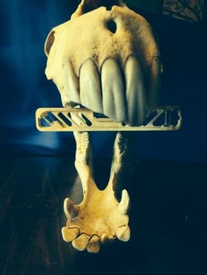
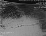
When deciduous teeth are lost at an early age due to trauma, there is considerable difference of opinion as to the eventual outcome. It has been stated that if the injury is more than six months prior to the expected eruption, the presence and viability of normal permanent dentition is in doubt. But, it is obvious from this radiograph that the 401 and 301 are already formed at ten months of age – more than a year and a half prior to normal eruption. The problem that remains is that with the loss of the central deciduous dentition, subsequent drifting of adjoining teeth will block the eruption pathway. Here is shown a method to hopefully prevent this drift. A small depression was created in the mesial surface of the 2’s and a slightly overlong 1/8 in. stainless rod was wedged into place. Further stability and protection was added with PMMA at both ends. We will await the eventual outcome of this case!
Richard O. Miller, DVM, EDPA/C
23411 Via Alondra
Coto de Caza, CA 92679
Case of the Month:
Open Pulps
By: Richard O. Miller DVM, EDPA/C
July 2017
OPEN PULPS Attention was first drawn to this 209 due to the fracture that had not been noted in two previous dental procedures. On closer inspection it was noted that all five pulps are open and subsequently probed deeply. Radiographs are not available as the horse is in a very remote region. Philosophically, one might consider that if the tooth is completely devitalized, is it more brittle and therefore caused the fracture or are the two lesions totally unrelated (?) From a practical standpoint one must consider a course of action. The most conservative approach would be to take the tooth out of occlusion since there are no obvious clinical symptoms. As is often the case, finances are quite limited but if this were taken as a charity situation, than the more direct, aggressive and proper decision would be to extract the tooth. That brings into play other issues: is their sufficient skill and instrumentation available to accomplish this in a remote region where follow up and aftercare may not be available? Will subsequent corrective procedures be scheduled to allow caudal/rostral drift? All of these variables should be discussed with the client with the best interest of the patient in mind.
Richard O. Miller, DVM, EDPA/C
23411 Via Alondra
Coto de Caza, CA 92679
Case of the Month:
Supernumeraries
By: Richard O. Miller DVM, EDPA/C
June 2017
SUPERNUMERARIES:
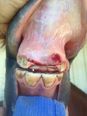
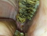
This condition is not rare but is considered uncommon. Although extra teeth (polydontia) may occur anywhere, this scenario is the most commonly missed. It has occurred that surgeons and radiologists, as well as practitioners, did not count past 11. Often this situation can be managed by yearly reduction but the picture shows a retromolar ulcer that is likely caused by the protuberant 212. In this case, and that shown on the skull of another horse, the angle of eruption has created a major periodontal lesion that prompted the decision to extract the supernumerary. This case was contributed by certified dental provider, Juston Hutchinson from Oklahoma, in cooperation with his veterinarian.
Richard O. Miller, DVM, EDPA/C
23411 Via Alondra
Coto de Caza, CA 92679
Case of the Month:
Sialolyths
By: Richard O. Miller DVM, EDPA/C
May 2017
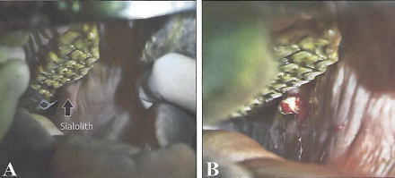
Sialolyths, or salivary duct stones have been mentioned before and have a similar cause as enterolyths –the stones that form in the equine colon. Usually an irritating object is the inciting cause and mineral deposits build up in an attempt to isolate it. In the case of sialolyths, the object is likely to be a foxtail or other type of plant awn. The condition is uncommon if not rare and can occur in all of the salivary ducts. I have only seen it in the distal parotid duct, lateral to the upper 9’s. A few cases have been reported in the proximal segment of the duct – much closer to the gland. Those few cases cannot be reached orally and require a transcutaneous approach. In the case shown it would be inappropriate to use such an approach, as there is a chance for skin infection as well as some risk to blood vessels and nerves in the area and a possibility of fistula formation. The transoral approach is easily reached with a # 12 scalpel blade clamped to a long intestinal forceps. There is typically no after care. In this particular case, considerable atrophy of the parotid gland was noted. Whether function will return is unknown. (photos courtesy of Nicholas Carlson, DVM of Salinas, Ca. and Tiago Silvino, DVM of Porto Alegre, Brazil )
Richard O. Miller, DVM, EDPA/C
23411 Via Alondra
Coto de Caza, CA 92679
Case of the Month:
Radiographs are a Diagnotic Aid
By: Richard O. Miller DVM, EDPA/C
April 2017
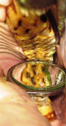
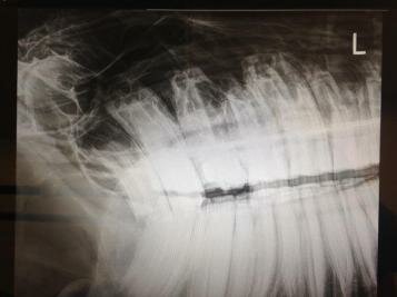
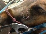
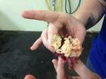
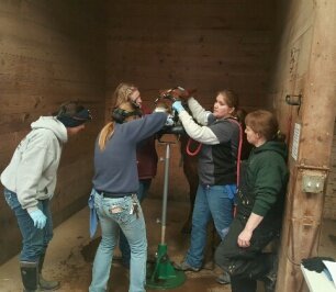
We have discussed in the past, the value of pre and postsurgical radiographs but what has not been emphasized is the fact that radiographs, with the best of interpretation, are only a diagnostic aid. From the radiograph shown, it is obvious that the 209 is affected. On intraoral exam with diligent and thorough spreading and manipulation, no progress was being made. The occlusal picture shows the attempt to drill and split the tooth in half and still no result, as all the excess tooth at the right was not visible. Finally, a standing osteotomy was performed and repulsion was successful.
Richard O. Miller, DVM, EDPA/C
23411 Via Alondra
Coto de Caza, CA 92679
Case of the Month:
Be Prepared
By: Richard O. Miller DVM, EDPA/C
March 2017
BE PREPARED. The Girl/Boy Scout Motto also applies to dentistry. This case was presented by certified EDP’s, Becca Greene and Natalie Hillman with the support of Susan Tavernier, DVM. The photos themselves offer a very interesting case study but my emphasis is on being prepared. We have all gone out on an ambulatory visit to do what we thought would be a routine maintenance day, only to be confronted by an unexpected fracture. In this case, preevaluation radiographs would seem to be unnecessary and how difficult can this 307 saggital fracture be? But, very wisely, Natalie deferred the extraction in favor of joining her full team in a warm clinical environment with all the necessary personnel, instrumentation and aftercare that might be required. And, as can be seen, from the 17 fragments that were removed, both from an intraoral as well as an extraoral approach, this was not a routine case. These fractures are seldom emergencies and most often showing no symptoms, so planning the actual procedure is usually the best course.
Richard O. Miller, DVM, EDPA/C
23411 Via Alondra
Coto de Caza, CA 92679
Case of the Month:
Zebra Skulls
By: Richard O. Miller DVM, EDPA/C
January 2017
What we would consider to be major ETR’s does not seem to be an issue but these specimens are young. Calculus formation on the canines at a young age is unexplained in my thoughts but this is such a small sample. Wolf teeth appear to be in contact with the rostral tip of the lower 06 ramps but, again, this might be only a factor in performance situations.
The disparity of width in the upper and lower incisor arcades does not seem to have caused anything more than slight upper caudal prominences on the upper 03’s. These images demonstrate the true angle of spee. NOT the ones occasionally published that are actually dominant upper 10’s that have compromised the opposing lower arcades. Thank you Graeme and Katherine Ros, DVM, for so much food for thought.
Richard O. Miller, DVM, EDPA/C
23411 Via Alondra
Coto de Caza, CA 92679
In order to speed up download times and yet still give our members and others access to these cases, we have provided them here. Simply click on the link above and download. It is in Microsoft Word. If you have any questions, or problems, please contact
Jan-Oct2013 Case of Month.doc
Microsoft Word document [849.5 KB]
In order to speed up download times and yet still give our members and others access to these cases, we have provided them here. Simply click on the link above and download. It is in Microsoft Word. If you have any questions, or problems, please contact
Cases Nov 13-Dec 14.docx
Microsoft Word document [1.5 MB]
In order to speed up download times and yet still give our members and others access to these cases, we have provided them here. Simply click on the link above and download. It is in Microsoft Word. If you have any questions, or problems, please contact
Cases Jan 15-Dec 15.docx
Microsoft Word document [1.8 MB]
In order to speed up download times and yet still give our members and others access to these cases, we have provided them here. Simply click on the link above and download. It is in Microsoft Word. If you have any questions, or problems, please contact
Cases Jan 16-Dec 16.docx
Microsoft Word document [2.1 MB]
